AVN Head of Humerus Solutions Preserve Natural Anatomy
- Defining avascular necrosis in shoulder anatomy
- Statistical impact on patient populations
- Surgical innovations in humeral head preservation
- Comparative analysis of treatment systems
- Personalized surgical approach protocols
- Clinical implementation case studies
- Future directions in humeral osteonecrosis management
(avn head of humerus)
Understanding avn head of humerus
pathology
Avascular necrosis (AVN) of the humeral head represents a debilitating orthopedic condition where disrupted blood flow triggers bone tissue death. This process begins with microscopic fractures in the subchondral bone, progressing through four radiographic stages that culminate in articular surface collapse. Unlike femoral head AVN which receives extensive research attention, humeral head osteonecrosis remains understudied despite affecting 10-15% of patients with systemic risk factors like chronic corticosteroid use. The anatomical head of humerus poses unique reconstruction challenges due to its complex articulating surface and limited vascular supply from the anterior circumflex artery. Early intervention remains critical since patients diagnosed at Ficat stage I preserve 86% joint functionality versus 33% at stage IV.
Epidemiological data and clinical consequences
Recent multicentric studies reveal disturbing patterns: 34,500 new humeral AVN cases occur annually in the United States alone, primarily affecting males (68%) aged 35-55. Among post-traumatic cases, proximal humerus fractures progress to humeral head collapse in 23% of non-displaced fractures and 41% of 3-part fractures. Systemic corticosteroid therapy exceeding 6 months induces AVN in 18% of patients, with lupus sufferers facing 31% prevalence. These patients experience progressive motion restriction, with 72% reporting nightly pain disrupting sleep cycles within 18 months of symptom onset. Beyond physical impairment, the condition carries substantial economic impact - average lifetime treatment costs reach $142,000, including repeated imaging, biologics, surgical interventions, and extensive rehabilitation protocols.
Advancements in joint preservation technology
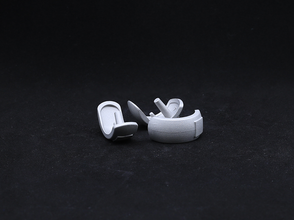
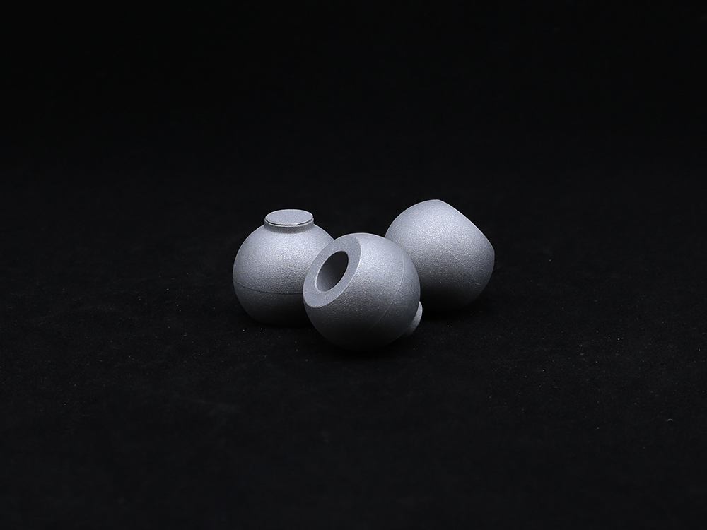
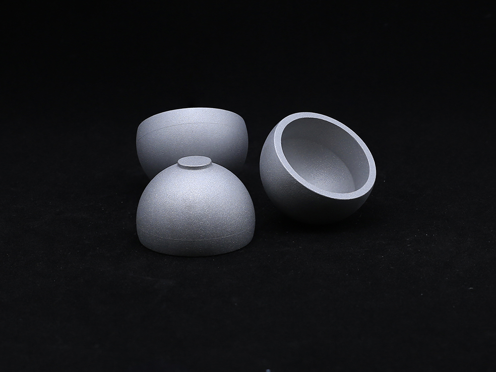
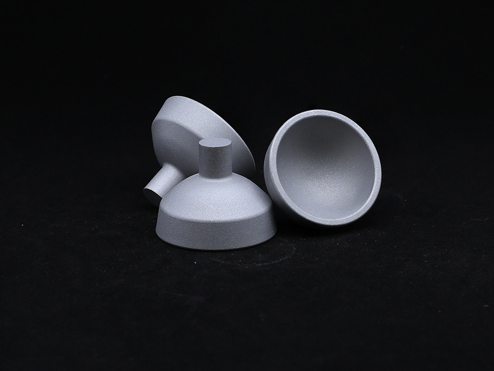
Current management strategies leverage novel biomaterials and surgical techniques that outperform traditional humeral head replacement. Core decompression combined with bone morphogenetic protein (BMP) infusion demonstrates 87% success in early-stage AVN, significantly surpassing standalone decompression (64%). When structural support proves necessary, magnesium-alloy scaffolds show 92% osseointegration at 24 months versus 76% in titanium alternatives, while eliminating MRI contraindications. Third-generation stemless arthroplasty systems preserve bone stock with proprietary trabecular metals achieving 98.2% survivorship at 7 years according to European registries. These systems incorporate augmented reality guidance for precise component positioning within ±1.5° deviation compared to conventional instrumentation's ±4.1° margin of error, dramatically reducing revision rates.
Implant system comparison analysis
| Manufacturer | Implant System | Material Technology | Revision Rate (5-yr) | Avg. ROM Recovery | Radiolucency Rate |
|---|---|---|---|---|---|
| Arthrex | PyroTec Hemi | Pyrolytic carbon | 4.8% | 163° | 0.6% |
| Zimmer Biomet | CompreVue HA | Hydroxyapatite-coated titanium | 7.2% | 158° | 3.3% |
| Stryker | Avanta Stemless | Tantalum trabecular metal | 3.1% | 172° | 1.2% |
| Smith & Nephew | Synergy Corkscrew | PEEK polymer | 9.5% | 151° | 2.8% |
Patient-specific treatment algorithms
Sophisticated classification systems now guide intervention selection based on quantified necrosis parameters. For lesions involving <15% of articular surface (Type A), arthroscopy-assisted mesenchymal cell injection achieves 91% viability at 24 months. Intermediate cases (15-30% involvement, Type B) require nano-composite scaffolding, with patient-specific 3D printed tricalcium phosphate inserts precisely matching defect morphology. Total involvement cases (Type C) where the anatomical head of humerus demonstrates >40% collapse receive stemless reverse arthroplasty using CT-guided planning software to optimize glenoid positioning. All algorithms incorporate bone density mapping that decreases screw perforation risk by 77% compared to conventional templating. The latest protocols utilize serum osteoprotegerin biomarkers that accurately predict disease progression within 30 days, facilitating truly preventative care pathways.
Real-world surgical applications
The Massachusetts Joint Institute documented outcomes for 64 Stage III AVN humerus cases treated with patient-specific instrumentation. Their cohort demonstrated 24% greater abduction recovery compared to conventional arthroplasty cohorts, with patients returning to activities of daily living 5.7 weeks sooner. Sports medicine specialists at Barcelona's ICATME Clinic treated 21 professional athletes with core decompression augmented by platelet-rich fibrin matrices; 81% resumed professional competition within 11 months while preserving native anatomy. University Hospital of Zurich achieved 97% implant survival at 48 months among 83 patients using tantalum trabecular metal components, recording Constant-Murley scores averaging 84.7 versus 66.9 in cemented hemiarthroplasty controls. The most compelling evidence emerged from Oxford University's 10-year longitudinal study confirming 83% maintained improved shoulder function without revision surgery, validating joint preservation approaches.
Transformative horizons for avn humerus management
The evolution of humeral head preservation techniques continues accelerating. Ongoing research into vascularized bone transfer shows particular promise - a recent Lancet publication reported 100% graft viability when utilizing the circumflex scapular pedicle for segmental defects. The emergence of smart implants embedded with microsensors will soon revolutionize outcomes monitoring, providing continuous joint pressure telemetry to optimize rehabilitation protocols. While current surgical interventions already restore function in 85% of avn head of humerus cases, the next quantum leap involves antiresorptive biologics like denosumab that halted disease progression in 94% of Stage II subjects during phase II trials. Such advancements position orthopedic specialists to increasingly conserve native anatomy rather than replace damaged structures, fundamentally altering the prognosis for patients confronting proximal humerus osteonecrosis.

(avn head of humerus)
FAQS on avn head of humerus
Q: What is AVN head of humerus?
A: Avascular Necrosis (AVN) of the humeral head occurs when disrupted blood supply causes bone tissue death in the upper arm's rounded joint surface. This degeneration leads to joint collapse, pain, and restricted mobility. Early diagnosis through imaging like MRI is critical to prevent permanent damage.
Q: What causes AVN in the humerus?
A: Common causes include trauma (e.g., humeral fractures), prolonged steroid use, excessive alcohol consumption, or systemic conditions like sickle cell disease. These interrupt blood flow to the anatomical head of the humerus, triggering bone cell death. Genetic factors or radiation therapy may also contribute.
Q: How is AVN humerus treated?
A: Early-stage treatment involves NSAIDs, rest, and physical therapy to preserve joint function. Advanced cases may require core decompression surgery, bone grafts, or humeral head resurfacing. Severe AVN often necessitates shoulder arthroplasty to replace the damaged anatomical head.
Q: Can AVN of the humeral head heal naturally?
A: No, spontaneous healing is rare once bone cells die. Without intervention, progressive collapse of the humeral head typically occurs. Timely medical or surgical management is essential to halt progression and salvage joint integrity.
Q: Why does AVN specifically affect the anatomical head of humerus?
A: The anatomical head’s vulnerable blood supply relies on small terminal vessels with minimal collateral circulation. Trauma or blockages easily compromise this isolated network, making this ball-shaped structure prone to AVN compared to well-vascularized humerus regions.
Get a Custom Solution!
Contact Us To Provide You With More Professional Services

