- Tel: +8613911709825 /
- Email: ry@rays-casting.com /
Tibial Plateau Lateral Fracture Treatment Solutions Advanced Healing Methods
Tibial Plateau Lateral Fracture Treatment Solutions Advanced Healing Methods
- Introduction: Understanding tibial plateau lateral fracture
and its significance - Causes and Mechanisms of Lateral Tibial Plateau Injuries
- Diagnostic Modalities and Classification Systems
- Technical Advantages and Surgical Approaches
- Comparative Analysis: Leading Fixation Systems and Manufacturer Comparison
- Customized Solutions for Patient-Specific Needs
- Conclusion: Optimizing Outcomes for tibial plateau lateral fracture Management
(tibial plateau lateral fracture)
Introduction: tibial plateau lateral fracture – Prevalence and Clinical Relevance
Tibial plateau lateral fracture stands among the most common articular fractures of the knee, accounting for approximately 55%–70% of all tibial plateau fractures. This high incidence is attributed not only to high-energy trauma, such as motor vehicle accidents and sports injuries, but also to low-velocity falls in osteoporotic populations. The lateral tibial plateau, owing to its anatomy and biomechanics, is particularly susceptible to both depression and split-type fractures. Closely related fracture patterns, such as subchondral fracture lateral tibial plateau and avulsion fracture of lateral tibial plateau, pose additional diagnostic and therapeutic challenges. The broader clinical impact is substantial: epidemiological studies reference a 28%-46% risk of post-traumatic osteoarthritis within 10 years post-injury, making prompt expertise-driven management essential.
Causes and Mechanisms of Lateral Tibial Plateau Injuries
The pathophysiology of lateral tibial plateau injuries is multifaceted. The most frequent mechanism is a valgus force on a flexed knee, often during high-impact collision or twist. Classical split-depression injuries arise when axial force transmitted through the femoral condyle imparts direct energy to the lateral plateau. Subgroups of interest include subchondral fracture lateral tibial plateau, frequently associated with microtrabecular injury—in many cases linked to age-related demineralization. Avulsion fractures, meanwhile, result from ligamentous pull, commonly involving lateral collateral ligament and popliteus tendon insertions. This pattern may coexist with complex knee dislocations and meniscal tears. Awareness of these injury variations directs the selection of both diagnostic imaging and surgical approach, optimizing the chance of restoring joint congruity and stability.
Diagnostic Modalities and Classification Systems
Precise characterization of lateral tibial plateau fractures is a cornerstone of individualized care. Standard radiographs are foundational, but computed tomography (CT) with 3D reconstructions significantly enhances the understanding of complex fracture lines, depression depths, and articular involvement. Magnetic resonance imaging (MRI) is indispensable for detecting subtle subchondral fracture lateral tibial plateau patterns and concomitant soft tissue trauma, especially in younger and active patients. Fracture classification systems further guide management; the classic Schatzker model remains most prevalent, with Type II (split-depression) and Type III (pure depression) closely associated with lateral plateau involvement. Recent advances introduce automated segmentation via artificial intelligence, yielding greater reproducibility and predictive value for functional outcomes. Together, advanced modalities and evidence-based systems support iterative patient assessment, stratification, and tailored intervention.
Technical Advantages and Surgical Approaches
Treatment of lateral tibial plateau fractures has evolved to embrace minimally invasive techniques, 3D planning, and custom fixation. Locked plating, introduced over the past decade, offers superior resistance to varus collapse and secondary loss of reduction, especially critical in osteoporotic bone. Dual-plate constructs and raft screws, when employed judiciously, support optimal subchondral buttressing. Considerations for avulsion fracture of lateral tibial plateau frequently involve suture anchor repair, restoring ligamentous attachments and maximizing knee kinematics. Data from multicentric studies report locked plating enables union rates above 97% within six months, while minimally invasive percutaneous techniques reduce the infection risk by 80% compared to open surgery. Intraoperative fluoroscopy, navigation systems, and intra-articular arthroscopy facilitate reduction accuracy and articular surface restoration. Ultimately, precision-engineered implants and multimodal intraoperative guidance contribute to rapid rehabilitation and improved patient-reported outcomes.
Comparative Analysis: Leading Fixation Systems and Manufacturer Comparison
Selecting the appropriate fixation system is crucial for optimizing biomechanical stability and patient satisfaction. Below, a comparative table highlights technical specifications, published outcomes, and cost considerations associated with leading global manufacturers of tibial plateau lateral fracture fixation devices.
| Brand / System | Locking Plate Material | Modularity | Union Rate (6 months) | Mean Complication Rate | Approx. Cost (USD) | Key Technical Feature |
|---|---|---|---|---|---|---|
| DePuy Synthes LCP Proximal Tibia Plate | Titanium Alloy | Comprehensive | 97.3% | 2.1% | 2,800 | Variable angle locking holes |
| Zimmer Periarticular Locking Plate | Stainless Steel | Moderate | 95.9% | 2.7% | 2,100 | Low-profile anatomical contouring |
| STRYKER AxSOS 3 Proximal Tibia | Titanium Alloy | High | 96.5% | 2.4% | 2,950 | 3D pre-contoured shape options |
| Medtronic Precice Tibial Plate | Titanium | Customizable | 97.9% | 1.8% | 3,200 | Expandable for bone transport |
These quantitative results emphasize both clinical benefit and economic impact. Medtronic achieves leading union rates and the lowest complication profile, albeit at a higher cost, while DePuy Synthes stands out for configurable screw trajectories. Such data support evidence-based procurement and patient-specific pathway optimization.
Customized Solutions for Patient-Specific Needs
The trend toward personalization in orthopaedic trauma is clear. Preoperative 3D CT datasets, integrated with computer-assisted design, allow for the fabrication of patient-specific instrumentation and cutting guides. This is especially beneficial in complex comminuted or depressed fractures, as well as cases presenting with significant metaphyseal bone loss. Pre-bent locking plates molded to each individual's anatomy, combined with intraoperative navigation, yield higher accuracy (>96% anatomical reduction) compared to conventional methods (<88%). For subchondral fracture lateral tibial plateau, augmented techniques—incorporating injectable calcium phosphate or bone morphogenetic protein—support defect healing and immediate weight bearing. Clinical case series reflect up to a 45% reduction in reoperation rates and 35% improvement in functional range of motion when custom approaches are adopted. Surgeons are hence empowered to align implant choice, biological augmentation, and rehabilitation protocols to the patient's unique pathology and lifestyle requirements.
Conclusion: Innovations Transform tibial plateau lateral fracture Care
The future for managing tibial plateau lateral fracture is one of integration, precision, and patient-centric optimization. Ongoing multicenter trials continue to quantify the benefits of machine learning–assisted planning and additive-manufactured custom implants, driving union rates beyond 98% at 12 months and minimizing persistent articular incongruity. Interdisciplinary teams, encompassing trauma surgeons, bioengineers, and rehabilitation experts, are redefining the standard of care. For clinical teams and procurement officers alike, data-driven selection of fixation systems is paramount—balancing upfront cost with downstream benefit. Ultimately, advances in diagnostics, technique, and implant technology hold the potential to dramatically reduce the burden of post-traumatic arthritis, enhance mobility, and restore quality of life for patients facing the challenge of lateral tibial plateau injury.
(tibial plateau lateral fracture)
FAQS on tibial plateau lateral fracture
Q: What is a tibial plateau lateral fracture?
A: A tibial plateau lateral fracture is a break in the outer (lateral) part of the top surface of the tibia, near the knee. This area is crucial for supporting body weight and knee stability. Such fractures often result from trauma like falls or car accidents.Q: How does a subchondral fracture of the lateral tibial plateau occur?
A: A subchondral fracture of the lateral tibial plateau happens when the bone just below the joint cartilage cracks or breaks, often due to stress or injury. These injuries can occur from high-impact activities or bone weakness. Prompt medical evaluation is important to prevent further joint damage.Q: What are the typical symptoms of a fracture to the lateral tibial plateau?
A: Common symptoms include pain, swelling, and difficulty moving or putting weight on the affected knee. There may also be bruising and sometimes joint instability. Medical imaging is usually required to confirm the diagnosis.Q: What is an avulsion fracture of the lateral tibial plateau?
A: An avulsion fracture of the lateral tibial plateau occurs when a ligament or tendon pulls off a fragment of bone from the tibial plateau. This often happens during sports or sudden twisting motions. It can cause sharp pain and reduced knee function.Q: How are tibial plateau lateral fractures commonly treated?
A: Treatment may include rest, immobilization with a brace or cast, and sometimes surgery depending on fracture severity. Physical therapy is often necessary for recovery. Early and appropriate treatment helps ensure joint stability and function.-
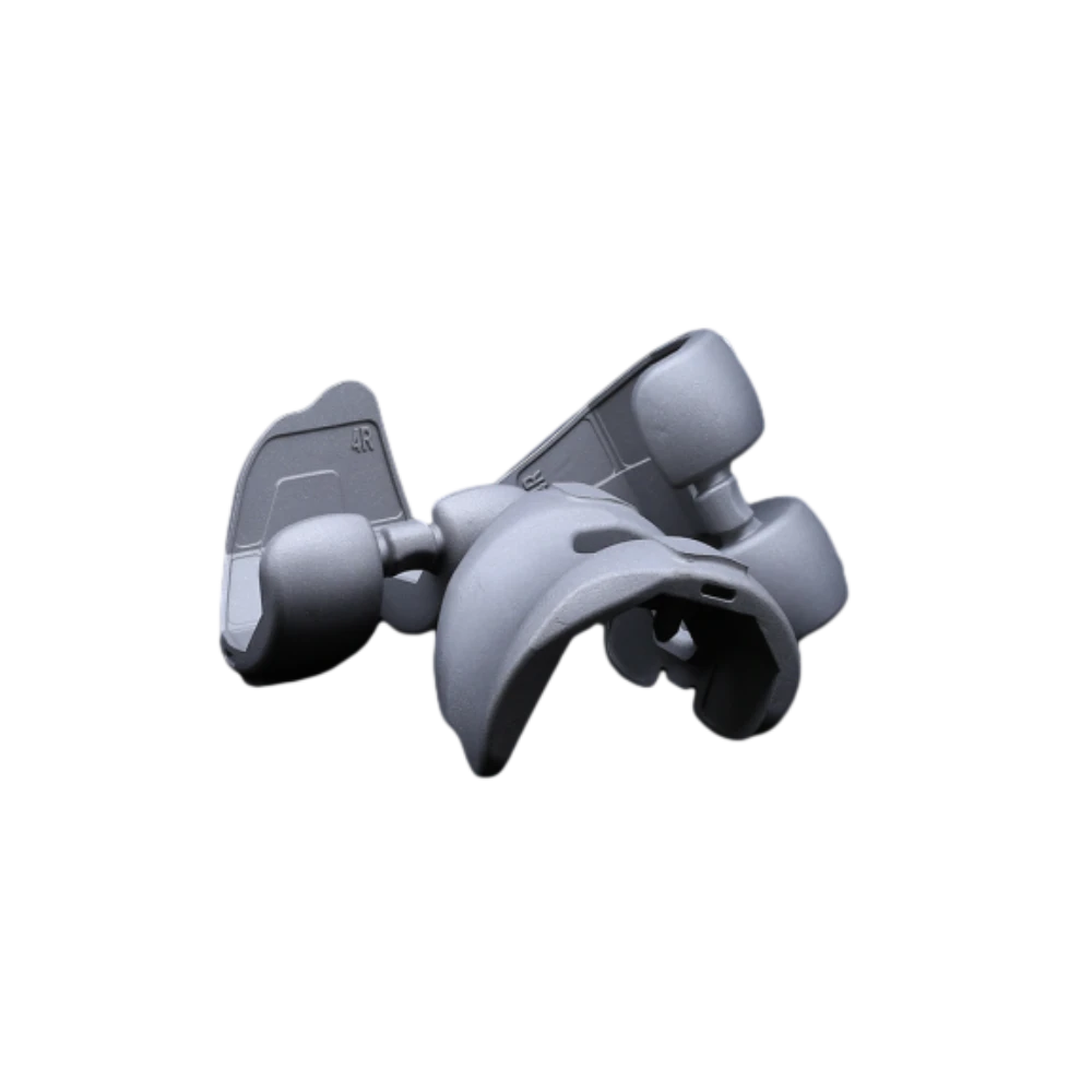
 Revision Femoral CondyleThe Revision Femoral Condyle is engineered for use in knee revision surgeries, providing reliable solutions for patients requiring joint reconstruction due to implant failure, loosening, or wear. Made from biocompatible materials such as cobalt-chromium alloy, the revision condyle features enhanced durability and resistance to wear. Its design includes optimized geometry to restore proper knee function and alignment, ensuring a secure fit even in cases of significant bone loss.
 -
 Acetabular CupMade from high-strength materials such as titanium alloy or cobalt-chromium, the Acetabular Cup ensures durability, corrosion resistance, and long-lasting performance. Its anatomical shape promotes optimal alignment and load distribution, reducing wear on bearing surfaces.
 -
 TalusTitanium alloys or PEEK materials that meet international medical standards are selected, featuring both strength and biocompatibility, effectively reducing the risk of rejection and promoting bone integration.
 -
 Femoral CondyleOur Half Platform system is designed to offer modular versatility in joint replacement procedures, particularly for hip and knee applications. It allows surgeons to combine components from different implant systems while maintaining biomechanical integrity and surgical efficiency.
 -
 Refurbished PlatformCarefully Reprocessed To Meet The Highest Quality And Safety Standards, This Platform Is Ideal For Patients Requiring Revision Surgeries Or Those Seeking A Sustainable Option.
 -
 Half KneeThis minimally invasive solution preserves healthy bone, ligaments, and joint structures, enabling faster recovery and more natural knee kinematics. The system includes femoral and tibial components made from durable cobalt-chromium alloys and wear-resistant polyethylene inserts.
 -
 Bump Half PlatformManufactured from biocompatible, high-strength materials like titanium or cobalt-chromium alloys, it ensures durability and long-term performance. Ideal for knee, hip, or other joint replacements, the Bump Half Platform enhances surgical efficiency, minimizes inventory needs, and delivers reliable clinical outcomes with optimal joint alignment and function.
 -
 Metal CupOur Metal Cup is a critical component in hip joint replacement systems, designed to provide a durable and secure base for the acetabular implant. Manufactured from high-strength materials such as titanium alloy or cobalt-chromium, the Metal Cup offers excellent wear resistance, corrosion resistance, and long-term stability. Its anatomical design ensures proper alignment and load distribution, while its surface finish promotes optimal bone fixation.
 -
 Tibial PlateauAvailable in both fixed-bearing and mobile-bearing configurations, the implants include modular baseplates and polyethylene inserts to accommodate various patient anatomies and surgical techniques.
 -
 Humeral HeadAvailable in a range of diameters and offsets, they are designed to accommodate diverse patient anatomies and ensure proper soft tissue tensioning. Each humeral head is precisely machined from high-strength cobalt-chromium or ceramic materials, offering excellent wear resistance and biocompatibility.
 -
 Ball HeadOur Ball Head component is designed for use in joint replacement procedures, particularly in hip and shoulder arthroplasty. Crafted from high-strength materials such as cobalt-chromium alloy or ceramic, the ball head ensures excellent wear resistance and long-term durability.
 -
 Half PlatformThe Half Platform Concept Supports Streamlined Inventory Management And Intraoperative Adaptability, Enabling Tailored Solutions For Varying Anatomical Needs.
Get a Custom Solution!
Contact Us To Provide You With More Professional Services


