- Tel: +8613911709825 /
- Email: ry@rays-casting.com /

Femoral Condyle Conditions and Femoral Chondral Defects: From Diagnosis to Precision Orthopedic Solutions
Femoral Condyle Conditions and Femoral Chondral Defects: From Diagnosis to Precision Orthopedic Solutions
The femoral condyle plays an essential role in knee joint movement, load distribution, and overall joint stability. Located at the lower end of the femur, the medial and lateral femoral condyles articulate with the tibial plateau and interact with the patella to support daily activities such as walking, running, squatting, and climbing stairs. When this region experiences structural damage, cartilage degeneration, or compromised blood supply, patients may develop persistent knee pain and restricted mobility.
For orthopedic specialists, medical device manufacturers, and healthcare providers, understanding femoral condyle conditions is important for selecting appropriate diagnostic methods and treatment strategies. Conditions such as avascular necrosis (AVN) and femoral chondral defects require careful assessment of bone integrity, cartilage condition, lesion location, and the patient's functional needs. Advances in imaging, surgical planning, and orthopedic implant manufacturing continue to support more individualized approaches to knee joint preservation and reconstruction.

Understanding Femoral Condyle Anatomy and Its Role in Knee Joint Function
The femoral condyle consists of two rounded structures at the distal end of the femur. The medial femoral condyle is located on the inner side of the knee, while the lateral femoral condyle sits on the outer side. Together, they create the articular surfaces that help the knee move smoothly while transferring forces between the femur and tibia.
These surfaces are covered by articular cartilage, a specialized tissue that reduces friction and distributes pressure during movement. Healthy cartilage is particularly important because it allows the joint to withstand repetitive loading without excessive wear. Beneath the cartilage, subchondral bone provides structural support and helps maintain the shape of the joint surface.
Damage to the femoral condyle can result from acute trauma, repetitive mechanical stress, degenerative joint disease, or impaired blood circulation. Depending on the severity and location of the lesion, patients may experience pain during weight-bearing activities, swelling, stiffness, or reduced range of motion. Some conditions develop gradually, while others become symptomatic after an injury.
Diagnostic evaluation generally combines a clinical examination with imaging studies. X-rays can reveal changes in bone structure, joint space, and alignment. Magnetic resonance imaging (MRI) provides more detailed information about cartilage, bone marrow abnormalities, and soft tissue involvement. Computed tomography may also be useful when a more precise assessment of bone geometry is required for surgical planning.
For orthopedic treatment planning, identifying the exact location and extent of a lesion is essential. A small cartilage defect with preserved underlying bone may require a different approach from a large lesion involving both cartilage and subchondral bone. Accurate anatomical assessment helps clinicians evaluate available treatment options and determine whether joint-preserving procedures or reconstructive surgery may be appropriate.
How AVN Femoral Condyle Affects Bone Integrity and Treatment Planning
Avascular necrosis of the femoral condyle, commonly described as AVN femoral condyle, occurs when the blood supply to a region of bone becomes insufficient, resulting in bone tissue damage. The condition may affect the medial or lateral femoral condyle and can progress from early bone changes to subchondral fracture and collapse if the affected area deteriorates.
Patients may report deep knee pain that worsens with standing, walking, or other weight-bearing activities. In early stages, conventional radiographs may appear relatively normal, making MRI particularly valuable for identifying bone marrow changes and areas of suspected necrosis. As the disease progresses, imaging may reveal a defined lesion, changes in the subchondral bone, or flattening of the articular surface.
Several factors can influence the development of avascular necrosis, including corticosteroid exposure, excessive alcohol consumption, certain systemic conditions, and local vascular or mechanical factors. However, the cause is not always identifiable. Clinicians must distinguish AVN from other causes of knee pain, including subchondral insufficiency fractures, osteoarthritis, and other bone lesions, because these conditions may require different management strategies.
Treatment depends on lesion size, location, disease stage, patient age, activity level, and the degree of joint surface damage. Early-stage cases may be managed with activity modification, appropriate pain management, protected weight-bearing, and specialist monitoring. Selected patients may be considered for joint-preserving procedures, such as core decompression or other interventions designed to address the affected bone.
When substantial bone collapse or advanced joint degeneration develops, reconstructive options may become necessary. Depending on the extent of damage, these may include procedures addressing the affected condyle or partial or total knee arthroplasty.
From a medical device manufacturing perspective, reconstructive treatment places considerable importance on anatomical accuracy, material properties, dimensional consistency, and quality assurance. Components intended for orthopedic applications must be manufactured according to applicable design specifications and validated processes. Close coordination between surgeons, engineers, and manufacturers helps ensure that device geometry and mechanical characteristics align with the intended clinical application.
Femoral Chondral Defects and Their Impact on Cartilage Preservation
A femoral chondral lesion refers to damage involving the articular cartilage covering the femoral condyle. Unlike injuries that primarily affect bone, a femoral chondral defect may initially involve the cartilage surface while the underlying bone remains intact. Depending on the cause and severity, the defect can range from superficial cartilage damage to a full-thickness lesion exposing the subchondral bone.
Common causes include sports injuries, direct trauma, repetitive joint loading, osteochondral injuries, and degenerative changes. Patients may experience knee pain, swelling after physical activity, stiffness, or catching sensations. However, symptoms vary considerably, and the presence of a cartilage defect does not always explain every symptom. Clinical findings must therefore be interpreted alongside imaging results and other possible sources of knee pain.
MRI is frequently used to assess cartilage thickness, lesion dimensions, associated bone marrow changes, and potential damage to surrounding structures. During treatment planning, clinicians consider whether the defect is focal or diffuse, whether the subchondral bone is involved, and whether other conditions, such as ligament instability or lower-limb malalignment, may contribute to abnormal loading.
Management strategies range from conservative care to surgical intervention. Nonoperative approaches may include activity modification, targeted rehabilitation, and symptom management. For selected focal cartilage defects, surgical options can include marrow stimulation techniques, osteochondral autograft or allograft transplantation, and cell-based cartilage repair procedures where clinically appropriate.
The choice of treatment depends on multiple factors, including lesion size, location, depth, patient activity demands, and the condition of the surrounding joint. A cartilage repair procedure may be unsuitable when widespread osteoarthritis or significant structural instability is present. Correcting contributing mechanical problems may also be important for protecting the repaired area and improving long-term joint function.
For orthopedic research and product development teams, femoral chondral defects highlight the need to understand the relationship between cartilage and underlying bone. Implants, graft-related instruments, and surgical tools must be designed around the requirements of their specific applications. Precise dimensions, suitable surface characteristics, reliable manufacturing processes, and thorough inspection all contribute to consistent product quality.
Choosing Orthopedic Manufacturing Solutions for Femoral Condyle Reconstruction
The treatment of femoral condyle injuries increasingly relies on collaboration among orthopedic surgeons, medical researchers, engineers, and specialized manufacturers. Although clinical decisions depend on individual patient assessments, high-quality orthopedic components can support surgical planning and reconstructive procedures when appropriately designed and indicated.
Manufacturing precision is especially important for components associated with the distal femur and knee joint. Small dimensional variations can affect fit, alignment, and the relationship between an implant and surrounding anatomical structures. Manufacturers therefore need controlled production processes, clearly defined specifications, and inspection procedures suited to the component's intended use.
Depending on the product and its design, manufacturing methods may include precision machining, casting, surface finishing, and other specialized processes. Material selection must consider the intended mechanical requirements, biocompatibility, corrosion resistance where relevant, and applicable medical device standards. Traceability, documentation, and process validation are also important elements of a reliable orthopedic supply chain.
For companies developing products related to femoral condyle reconstruction, cooperation with experienced manufacturing partners can help address design feasibility, dimensional tolerances, material requirements, and production consistency. Early communication between engineering and clinical teams can identify potential manufacturing challenges before they affect later development stages.
Ultimately, successful management of femoral condyle conditions requires an integrated understanding of bone health, cartilage preservation, joint mechanics, and appropriate treatment options. Whether clinicians are evaluating AVN femoral condyle or investigating a femoral chondral defect, accurate diagnosis remains the foundation of individualized care. For orthopedic manufacturers, translating detailed technical requirements into reliable, precisely produced components is an important part of supporting the continued development of knee reconstruction and joint-preservation solutions.
-
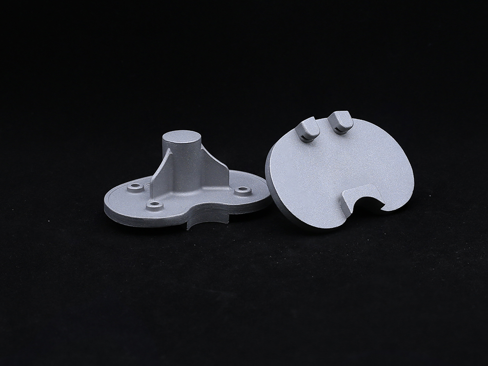 Refurbished PlatformCarefully Reprocessed To Meet The Highest Quality And Safety Standards, This Platform Is Ideal For Patients Requiring Revision Surgeries Or Those Seeking A Sustainable Option.
-
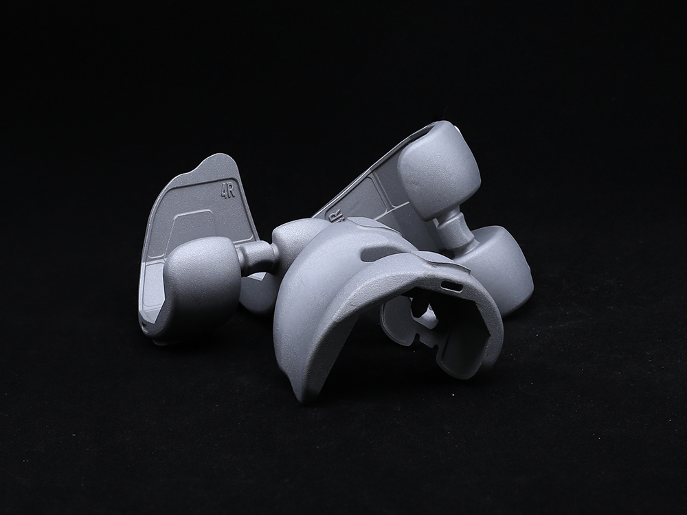 Femoral CondyleOur Half Platform system is designed to offer modular versatility in joint replacement procedures, particularly for hip and knee applications. It allows surgeons to combine components from different implant systems while maintaining biomechanical integrity and surgical efficiency.
-
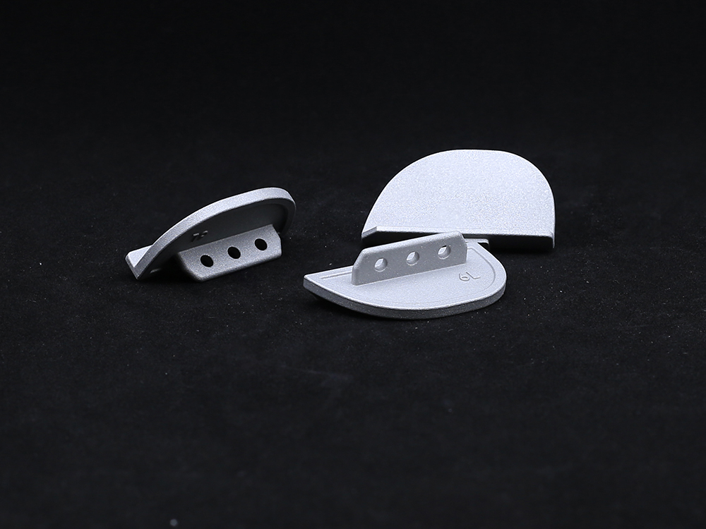 Unilateral Tibial PlateauThe unilateral tibial plateau is an orthopedic implant used in partial knee arthroplasty to replace the damaged medial or lateral tibial joint surface, thereby restoring function, relieving pain, and ensuring knee stability.
-
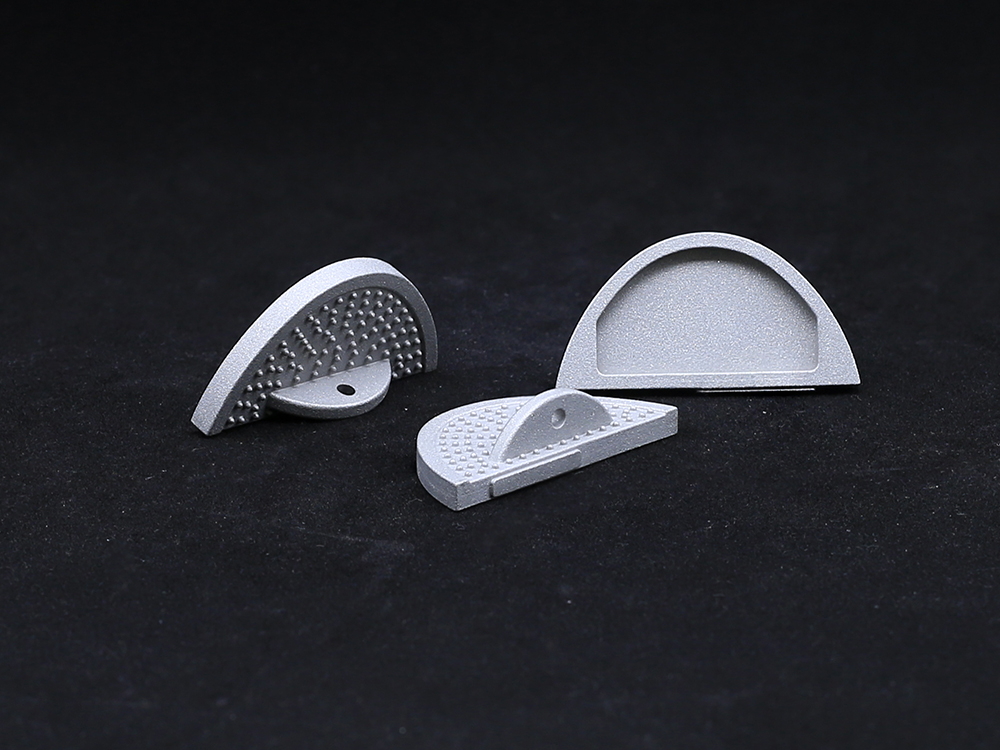 Bump Half PlatformManufactured from biocompatible, high-strength materials like titanium or cobalt-chromium alloys, it ensures durability and long-term performance. Ideal for knee, hip, or other joint replacements, the Bump Half Platform enhances surgical efficiency, minimizes inventory needs, and delivers reliable clinical outcomes with optimal joint alignment and function.
-
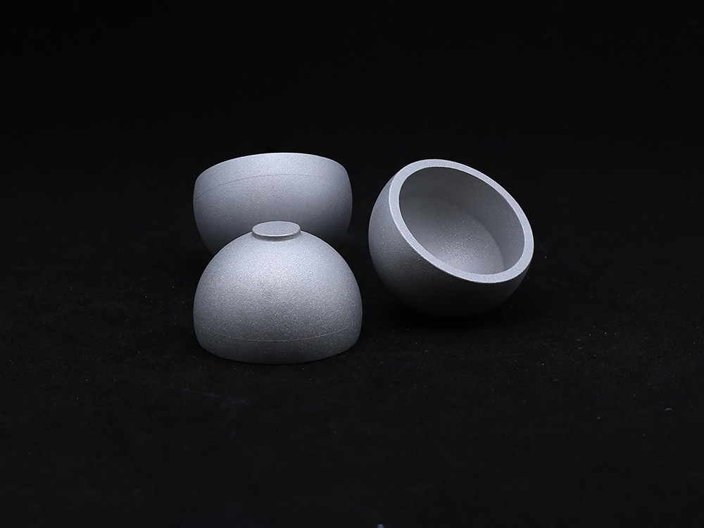 Metal CupOur Metal Cup is a critical component in hip joint replacement systems, designed to provide a durable and secure base for the acetabular implant. Manufactured from high-strength materials such as titanium alloy or cobalt-chromium, the Metal Cup offers excellent wear resistance, corrosion resistance, and long-term stability. Its anatomical design ensures proper alignment and load distribution, while its surface finish promotes optimal bone fixation.
-
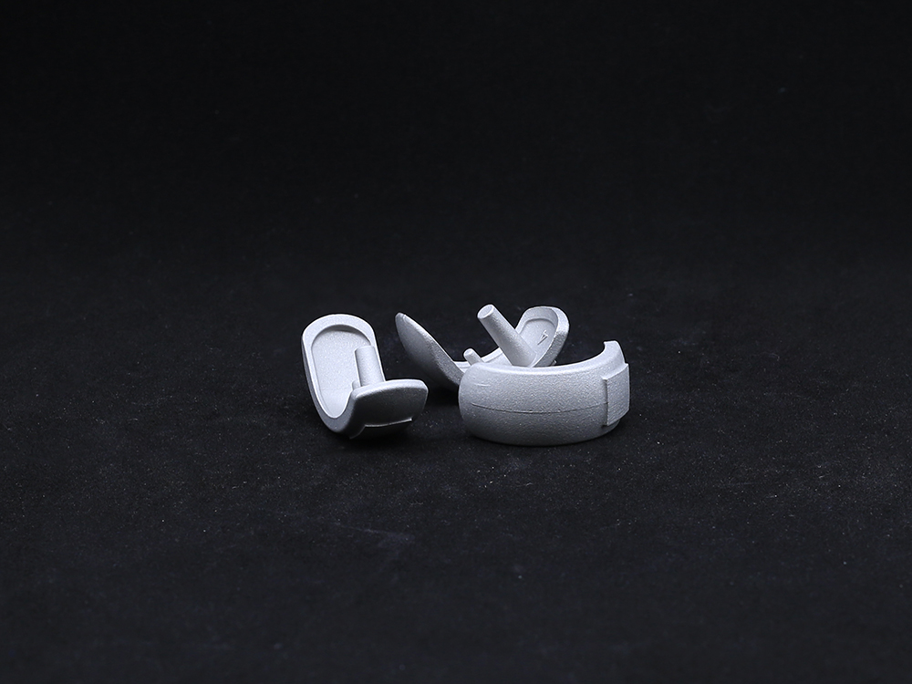 Half KneeThis minimally invasive solution preserves healthy bone, ligaments, and joint structures, enabling faster recovery and more natural knee kinematics. The system includes femoral and tibial components made from durable cobalt-chromium alloys and wear-resistant polyethylene inserts.
-
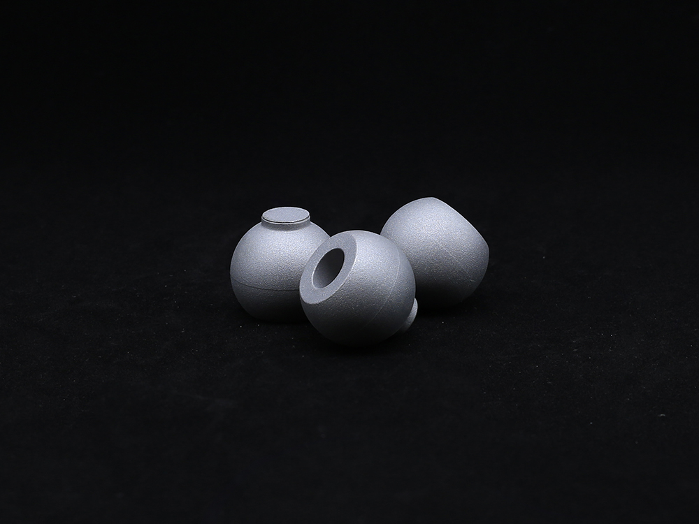 Ball HeadOur Ball Head component is designed for use in joint replacement procedures, particularly in hip and shoulder arthroplasty. Crafted from high-strength materials such as cobalt-chromium alloy or ceramic, the ball head ensures excellent wear resistance and long-term durability.
-
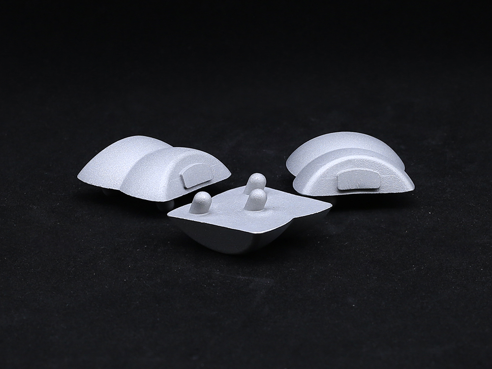 TalusTitanium alloys or PEEK materials that meet international medical standards are selected, featuring both strength and biocompatibility, effectively reducing the risk of rejection and promoting bone integration.
Get a Custom Solution!
Contact Us To Provide You With More Professional Services


