- Tel: +8613911709825 /
- Email: ry@rays-casting.com /

Fractured Femoral Condyle: Clinical Assessment, Implant Materials, and Precision Manufacturing for Knee Reconstruction
Fractured Femoral Condyle: Clinical Assessment, Implant Materials, and Precision Manufacturing for Knee Reconstruction
A fracture involving the femoral condyle can significantly affect knee joint stability, load distribution, and mobility. As part of the distal femur, the medial and lateral condyles support the articulation between the femur and tibia, while their smooth cartilage surfaces help maintain controlled movement. When a fracture extends into the joint, treatment must address not only bone healing but also articular surface congruity, limb alignment, and long-term joint function.
For orthopedic surgeons, medical device developers, and implant manufacturers, these injuries present interconnected clinical and engineering challenges. Fracture characteristics influence fixation strategies, material selection, and implant geometry, while manufacturing precision helps ensure that orthopedic components meet their specified design requirements. Understanding the relationship between anatomy, clinical needs, and production quality is essential for developing reliable solutions for complex knee injuries.

Clinical Challenges of a Fractured Femoral Condyle and Joint Surface Restoration
A fractured femoral condyle typically involves the rounded articular portion of the distal femur. These fractures may occur following high-energy trauma, such as vehicle collisions or serious falls, or lower-energy injuries in individuals with weakened bone. Some fractures affect a single condyle, while others extend across the distal femur or involve multiple joint surfaces.
The primary clinical concern is restoring the anatomical relationship between the femur and tibia. If the fracture produces displacement, depression, or rotation of the articular surface, abnormal contact pressure may develop within the knee. Inadequate restoration can contribute to stiffness, persistent pain, altered gait, and post-traumatic osteoarthritis. Associated soft tissue injuries, including ligament damage, may further complicate treatment.
Diagnostic assessment usually begins with physical examination and radiographic imaging. Standard X-rays help identify fracture location, displacement, and overall alignment. Computed tomography (CT) can provide a more detailed view of fracture lines, fragmented bone, and articular surface involvement, particularly when surgical planning requires precise anatomical information. MRI may be considered when cartilage, ligaments, or other soft tissue structures also need evaluation.
Treatment depends on fracture stability, displacement, bone quality, the condition of the surrounding soft tissues, and the patient's overall health. Selected nondisplaced fractures may be managed with immobilization and carefully supervised restrictions on weight-bearing. Displaced or unstable fractures frequently require surgical fixation to restore joint congruity and maintain alignment during healing. Depending on the fracture pattern, fixation options may include screws, plates, or combinations of these devices.
Rehabilitation is another important consideration. Weight-bearing progression and knee motion must be coordinated with fracture stability and healing. Excessively aggressive movement or loading may compromise fixation, whereas prolonged immobilization can contribute to joint stiffness. Individualized follow-up allows clinicians to monitor healing and adjust rehabilitation accordingly.
From a medical device perspective, these clinical requirements establish the foundation for implant design. Fixation components must accommodate the intended surgical approach, provide appropriate mechanical support, and remain compatible with the anatomical structures involved. Reliable treatment begins with accurate diagnosis and a fixation strategy tailored to the specific fracture rather than a one-size-fits-all approach.
Fractured Medial Femoral Condyle and the Importance of Implant Material Selection
A fractured medial femoral condyle affects the inner side of the distal femur and may disrupt an important load-bearing region of the knee. Depending on the injury mechanism, the fracture can involve the articular surface, extend into the metaphysis, or occur alongside ligament and soft tissue injuries. The degree of displacement and the stability of the remaining bone influence both treatment selection and implant requirements.
The medial condyle contributes to the distribution of forces transmitted through the knee during standing, walking, and other weight-bearing activities. When its articular contour is altered, restoring alignment and surface congruity becomes a major treatment objective. Surgeons assess the fracture configuration, bone stock, joint involvement, and any associated injuries before deciding whether conservative treatment or surgical fixation is appropriate.
For fractures requiring surgery, implant materials must balance mechanical performance with biological compatibility. Titanium alloys are widely used in orthopedic fixation devices because of their strength-to-weight characteristics, corrosion resistance, and established applications in medical implants. Stainless steel is another established option for selected orthopedic fixation products, offering different mechanical and manufacturing characteristics.
The appropriate material depends on the device design, loading conditions, intended duration of implantation, applicable standards, and clinical requirements. Material selection alone does not determine implant performance. Geometry, surface condition, connection design, screw placement, and the interaction between the implant and bone also influence the mechanical behavior of the fixation system.
Manufacturing controls are therefore essential. Machining operations must maintain specified dimensions and tolerances, while surface finishing should meet the requirements established for the particular device. Inspection may include dimensional verification, material traceability, and mechanical testing where required by the product's design and regulatory framework.
For orthopedic manufacturers, collaboration with clinicians and design engineers can help translate fracture-management requirements into practical component specifications. Reviewing loading conditions, anatomical constraints, and surgical handling requirements early in development can reduce design uncertainty and support more consistent production. Each finished device must still undergo the verification and validation activities appropriate to its intended medical application.
Hypoplastic Lateral Femoral Condyle and Anatomical Considerations in Knee Reconstruction
A hypoplastic lateral femoral condyle is an anatomical variation in which the outer femoral condyle is underdeveloped. Unlike an acute fracture, this condition relates to bone morphology rather than a break caused by trauma. Its clinical significance depends on the degree of underdevelopment, overall knee alignment, patellar tracking, and the presence of associated anatomical variations.
The lateral femoral condyle contributes to the shape and mechanics of the knee joint. Differences in its size or contour may influence the relationship between the femur, tibia, and patella. In certain patients, an underdeveloped condyle may be associated with altered joint mechanics or may be identified during evaluation for patellar instability, congenital conditions, or preoperative orthopedic planning. However, the finding does not automatically indicate that treatment is necessary.
Clinical assessment considers the patient's symptoms, physical examination findings, and relevant imaging. X-rays can help evaluate bone alignment and joint morphology, while CT may provide detailed information about osseous anatomy. MRI can help assess cartilage, ligaments, and other structures when associated abnormalities are suspected. The goal is to determine whether the anatomical variation contributes to the patient's symptoms or affects a planned surgical procedure.
For reconstructive planning, anatomical differences can create challenges in achieving appropriate alignment and implant fit. Standardized components may not accommodate every anatomical configuration equally well, making accurate preoperative measurements important. In selected procedures, surgeons may consider patient-specific planning or specialized implants when clinically justified.
These considerations also affect medical device engineering. Three-dimensional anatomical data can help designers evaluate component dimensions, contact geometry, and potential interference with surrounding structures. Computer-aided design and precision manufacturing may support the development of components that follow defined anatomical requirements, subject to clinical validation and regulatory approval.
It is important to distinguish an underdeveloped lateral condyle from a fractured condyle because the underlying problems and treatment objectives differ. A fracture generally requires assessment of bone stability and healing, whereas hypoplasia requires evaluation of anatomical morphology and its functional consequences. Accurate classification helps prevent inappropriate assumptions during diagnosis, surgical planning, and implant development.
Precision Manufacturing for Femoral Condyle Fixation and Reconstruction
The production of orthopedic devices for distal femoral injuries requires a coordinated approach to design, material control, dimensional accuracy, and quality management. Whether a component is intended for fracture fixation or a selected reconstructive procedure, its specifications must reflect the relevant clinical requirements and applicable medical device regulations.
Manufacturing methods vary according to the device. CNC machining can produce complex geometries and controlled dimensions, while specialized finishing processes help achieve specified surface characteristics. For devices with critical interfaces or complex contours, inspection plans should focus on the dimensions and features that affect assembly, fit, and mechanical performance.
Quality management extends beyond the machining stage. Material documentation, process controls, batch identification, inspection records, and traceability help establish consistency throughout production. Mechanical testing and other verification activities should be selected according to the design risks, intended use, and applicable standards. Manufacturers must also follow appropriate requirements for cleanliness, packaging, and sterilization when relevant to the finished product.
For healthcare providers and orthopedic companies sourcing medical components, supplier evaluation should consider manufacturing capabilities, documented quality systems, engineering communication, and experience with the required product category. Clear technical drawings, defined acceptance criteria, and early design reviews help align expectations between customers and production teams.
Ultimately, successful management of femoral condyle injuries depends on matching clinical needs with suitable treatment strategies and reliable medical technology. A fractured femoral condyle requires careful restoration of bone alignment and joint function, while a fractured medial femoral condyle may introduce specific considerations related to load-bearing anatomy and fixation. A hypoplastic lateral femoral condyle presents a different challenge centered on anatomical variation and individualized planning. By integrating clinical assessment, appropriate implant materials, and precision manufacturing, orthopedic teams can support the development of solutions designed around the functional demands of the knee joint.
-
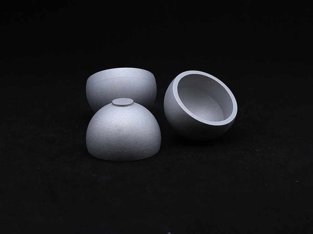 Metal CupOur Metal Cup is a critical component in hip joint replacement systems, designed to provide a durable and secure base for the acetabular implant. Manufactured from high-strength materials such as titanium alloy or cobalt-chromium, the Metal Cup offers excellent wear resistance, corrosion resistance, and long-term stability. Its anatomical design ensures proper alignment and load distribution, while its surface finish promotes optimal bone fixation.
-
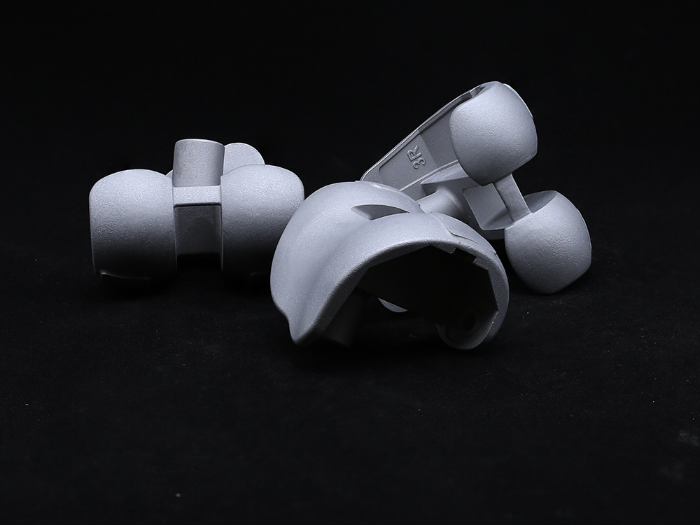 Revision Femoral CondyleThe Revision Femoral Condyle is engineered for use in knee revision surgeries, providing reliable solutions for patients requiring joint reconstruction due to implant failure, loosening, or wear. Made from biocompatible materials such as cobalt-chromium alloy, the revision condyle features enhanced durability and resistance to wear. Its design includes optimized geometry to restore proper knee function and alignment, ensuring a secure fit even in cases of significant bone loss.
-
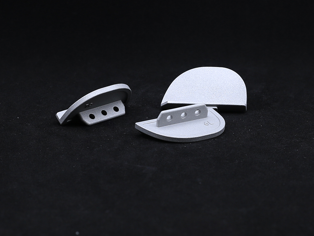 Unilateral Tibial PlateauThe unilateral tibial plateau is an orthopedic implant used in partial knee arthroplasty to replace the damaged medial or lateral tibial joint surface, thereby restoring function, relieving pain, and ensuring knee stability.
-
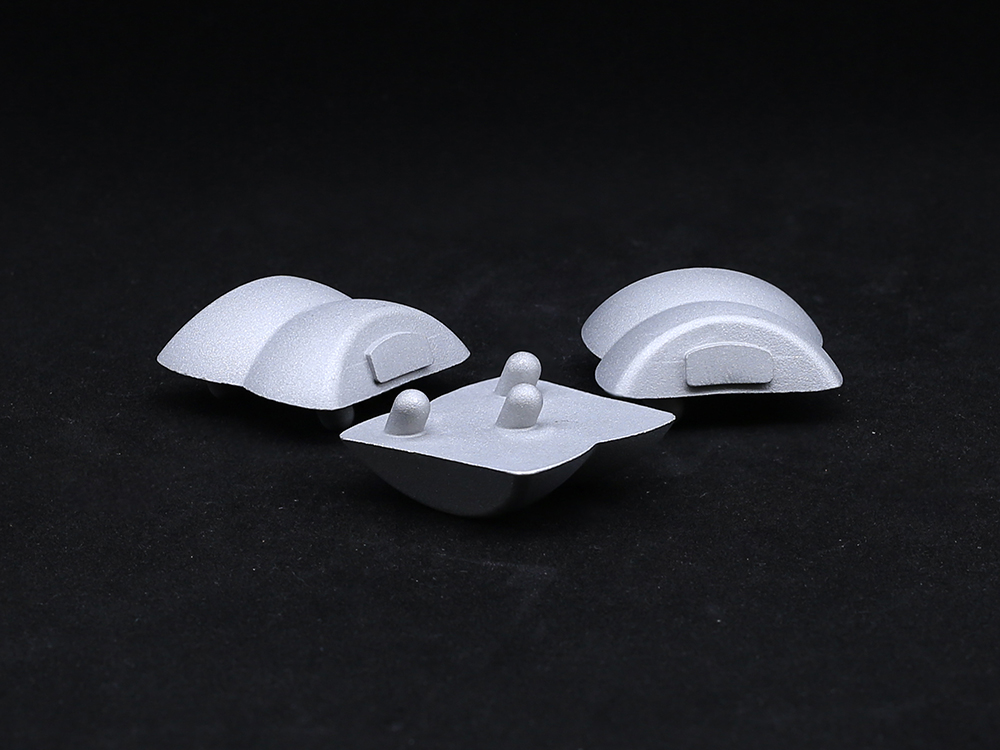 TalusTitanium alloys or PEEK materials that meet international medical standards are selected, featuring both strength and biocompatibility, effectively reducing the risk of rejection and promoting bone integration.
-
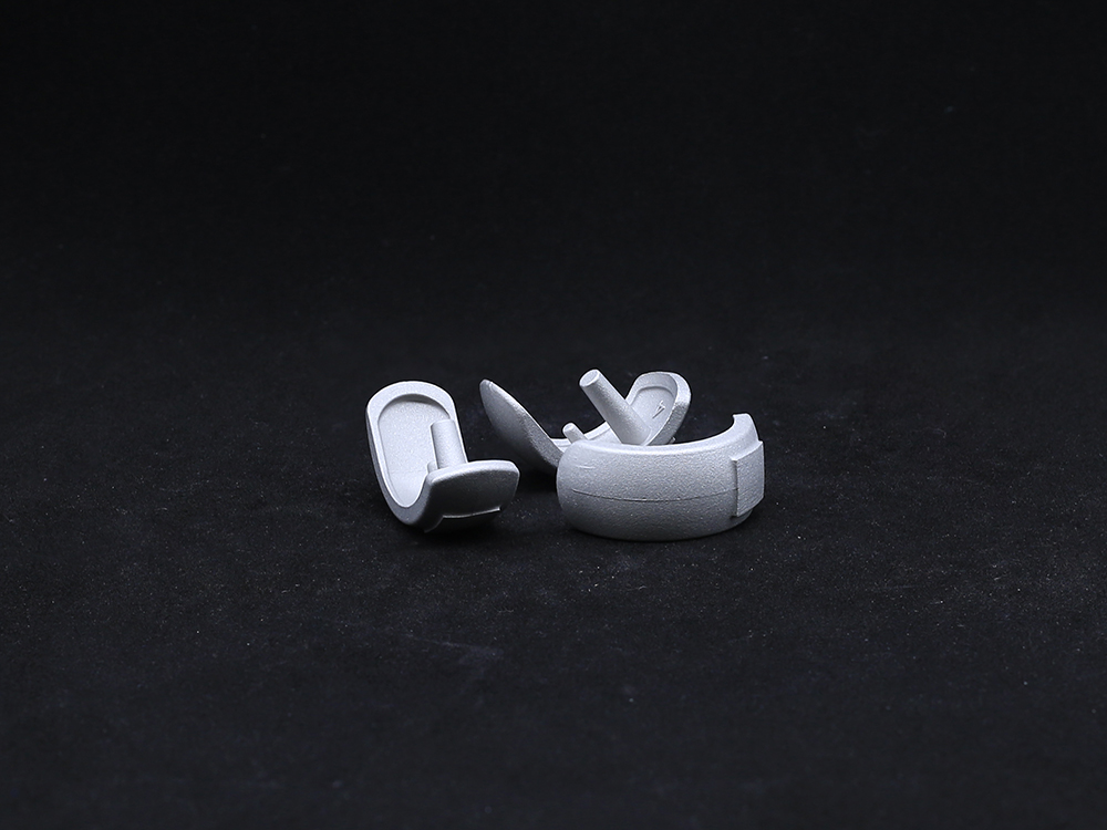 Half KneeThis minimally invasive solution preserves healthy bone, ligaments, and joint structures, enabling faster recovery and more natural knee kinematics. The system includes femoral and tibial components made from durable cobalt-chromium alloys and wear-resistant polyethylene inserts.
-
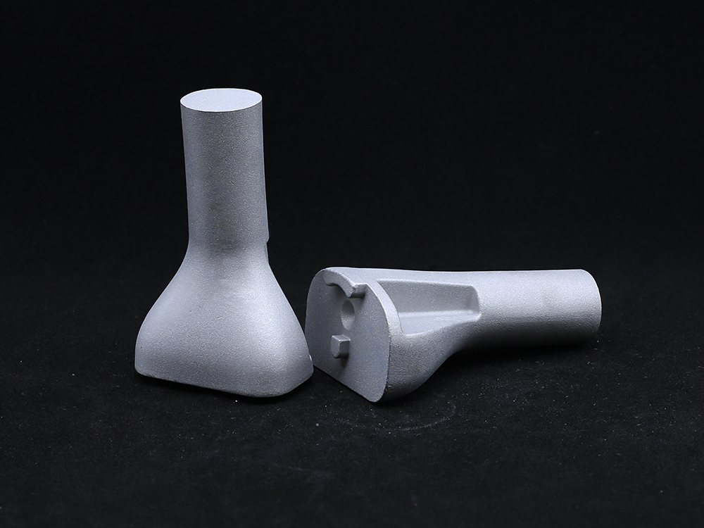 Tibial PlateauAvailable in both fixed-bearing and mobile-bearing configurations, the implants include modular baseplates and polyethylene inserts to accommodate various patient anatomies and surgical techniques.
-
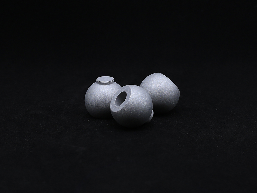 Ball HeadOur Ball Head component is designed for use in joint replacement procedures, particularly in hip and shoulder arthroplasty. Crafted from high-strength materials such as cobalt-chromium alloy or ceramic, the ball head ensures excellent wear resistance and long-term durability.
-
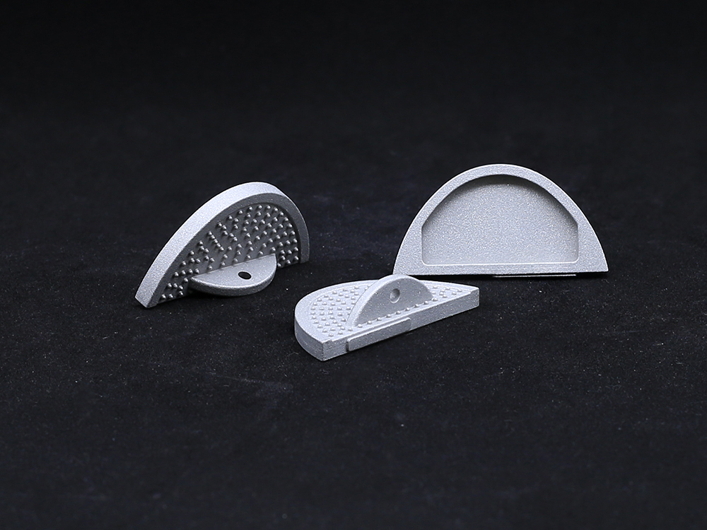 Bump Half PlatformManufactured from biocompatible, high-strength materials like titanium or cobalt-chromium alloys, it ensures durability and long-term performance. Ideal for knee, hip, or other joint replacements, the Bump Half Platform enhances surgical efficiency, minimizes inventory needs, and delivers reliable clinical outcomes with optimal joint alignment and function.
Get a Custom Solution!
Contact Us To Provide You With More Professional Services


