Central to Shoulder Function and Mobility
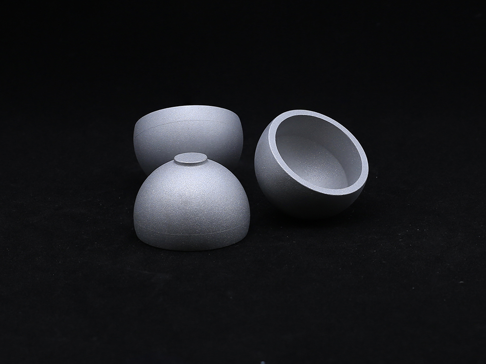
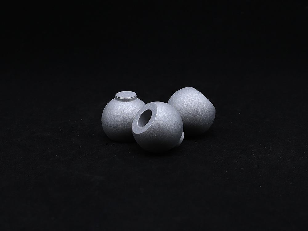
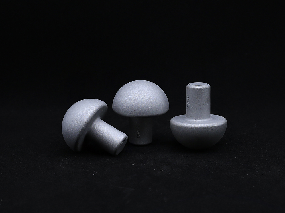
The humeral head is a vital anatomical structure that forms the ball portion of the shoulder’s ball-and-socket joint. Located at the top of the humerus (upper arm bone), the humeral head fits into the glenoid cavity of the scapula, creating the glenohumeral joint—the most mobile joint in the human body.
This spherical structure is covered with a smooth layer of articular cartilage, which allows it to glide effortlessly within the socket during arm movements. Because of its wide range of motion, the humeral head plays a critical role in everyday activities such as reaching, lifting, throwing, and rotating the arm. However, its mobility also makes it susceptible to wear and tear, traumatic injuries, and degenerative changes that may eventually require medical or surgical intervention.
Clinicians often evaluate the humeral head during shoulder exams or imaging studies, especially when patients report pain, stiffness, or loss of range of motion. Injuries like fractures, osteoarthritis, avascular necrosis, and labral tears frequently involve the humeral head and demand precise diagnosis for optimal treatment.
Cystic Changes in the Humeral Head: What Do They Mean?
When imaging reports reveal cystic changes humeral head, it usually refers to the presence of fluid-filled sacs or bone cysts within the humeral head's structure. These changes are commonly seen in MRI or CT scans and may indicate early signs of degeneration, stress injury, or osteoarthritis.
Cystic changes humeral head may be asymptomatic in some patients, discovered incidentally during imaging for unrelated issues. In other cases, they can be associated with deep, dull shoulder pain, weakness, or limited movement. These cystic formations typically occur in the subchondral bone beneath the cartilage, potentially signaling compromised bone strength or early degenerative joint disease.
While mild cystic changes humeral head can often be managed conservatively with physical therapy, anti-inflammatory medication, or injections, more advanced cases may require surgical intervention. In patients undergoing shoulder replacement or resurfacing, surgeons must evaluate the cystic bone quality to ensure implant stability and long-term success.
Recognizing and monitoring cystic changes humeral head early is crucial in preserving shoulder function and delaying the need for invasive procedures.
The Anatomical Head of Humerus: A Structural Marvel in Shoulder Design
The anatomical head of humerus is more than just a rounded bone—it is the engineering core of shoulder movement. It articulates with the glenoid fossa of the scapula and is part of a complex biomechanical system that supports the shoulder’s exceptional range of motion.
Unlike the surgical neck of the humerus, which lies just below, the anatomical head of humerus is located above the greater and lesser tubercles. It is hemispherical in shape and angled slightly upward and backward to optimize its articulation with the shoulder socket. Its smooth cartilage-covered surface allows it to rotate within the glenoid cavity with minimal friction, while nearby structures—like the rotator cuff, biceps tendon, and labrum—assist in stability and strength.
In orthopedic procedures such as hemiarthroplasty or total shoulder arthroplasty, replicating the natural geometry of the anatomical head of humerus is essential for restoring functional biomechanics. Prosthetic components are often modeled after the precise dimensions and orientation of the anatomical head of humerus to ensure natural shoulder movement after surgery.
Understanding this structure helps both patients and clinicians appreciate how even small abnormalities, such as flattening, collapse, or cystic changes humeral head, can lead to significant functional loss.
Injury and Degeneration of the Humeral Head: Signs You Shouldn't Ignore
Damage to the humeral head can result from trauma, overuse, or aging-related degeneration. Common conditions include osteoarthritis, rheumatoid arthritis, rotator cuff arthropathy, and avascular necrosis. In athletes and labor-intensive workers, repeated stress on the shoulder joint can cause microtrauma, leading to bone cysts or cartilage thinning.
When cystic changes humeral head or structural deformities in the anatomical head of humerus are present, patients may experience symptoms such as shoulder pain, catching or grinding sensations, restricted motion, and weakness. Over time, the joint space narrows, cartilage erodes, and the head may lose its round shape—affecting joint mechanics and leading to compensatory strain in surrounding tissues.
Advanced imaging, including MRI and CT scans, is crucial for assessing the extent of damage. If conservative treatments fail to provide relief, surgical options such as humeral head resurfacing, stemmed implants, or full shoulder arthroplasty may be recommended. The earlier structural deterioration is addressed, the better the chance of preserving joint integrity and function.
Restoring Shoulder Health: From Diagnosis to Personalized Treatment
With today’s medical advancements, patients suffering from cystic changes humeral head or abnormalities in the anatomical head of humerus have access to personalized, highly effective treatment pathways. These include non-invasive therapies such as guided physical rehabilitation, regenerative injections, and minimally invasive arthroscopic procedures.
In more advanced cases, orthopedic surgeons now use 3D imaging and custom-designed implants to restore the shape and motion of the humeral head. Whether it’s preserving bone structure with resurfacing techniques or replacing it with anatomically contoured prosthetics, the goal is always the same: restoring pain-free function and improving quality of life.
By understanding the role of the humeral head in joint mechanics and recognizing signs of early degeneration, patients and clinicians can work together to prevent irreversible damage. Regular checkups, early imaging, and prompt medical attention remain the best strategies for maintaining shoulder mobility for years to come.
Humeral head FAQs
What is the function of the humeral head in the shoulder joint?
The humeral head serves as the "ball" of the ball-and-socket shoulder joint. It allows for a wide range of motion in multiple directions, including flexion, extension, rotation, and circumduction. Covered with cartilage, it glides within the glenoid cavity to facilitate smooth, pain-free movement.
What are cystic changes in the humeral head and should I be concerned?
Cystic changes humeral head refer to small, fluid-filled sacs within the bone that can indicate early degenerative changes or stress-related bone damage. While some cysts are benign and asymptomatic, others may lead to joint instability or discomfort. If detected, your doctor may recommend monitoring, therapy, or further evaluation.
What is the difference between the anatomical head of humerus and surgical neck?
The anatomical head of humerus is the round, uppermost part that articulates with the shoulder socket, while the surgical neck lies just below the tubercles and is a common site for fractures. The anatomical head is critical for joint function and is replicated precisely in shoulder replacement surgeries.
Can cystic changes in the humeral head be reversed or treated non-surgically?
In some cases, yes. If cystic changes humeral head are mild and not causing symptoms, physical therapy, anti-inflammatory medications, or image-guided injections may help reduce progression. Regular follow-up imaging ensures that the condition is managed effectively without surgery.
When is surgery necessary for humeral head damage?
Surgery may be necessary when damage to the humeral head leads to chronic pain, joint instability, or impaired mobility that does not respond to conservative treatment. Procedures range from arthroscopic cleaning to full shoulder replacement, depending on the severity of damage and patient needs.
Get a Custom Solution!
Contact Us To Provide You With More Professional Services

