Understanding the Femoral Condyle
The femoral condyle is a fundamental structure located at the distal end of the femur, consisting of two prominent rounded projections: the medial and lateral condyles. These structures articulate with the tibial plateau, forming the core of the knee joint, which is essential for weight-bearing and movement. The condyles are covered by articular cartilage, a smooth, resilient tissue that enables the knee to move freely and absorb shocks during daily activities such as walking, running, and jumping.
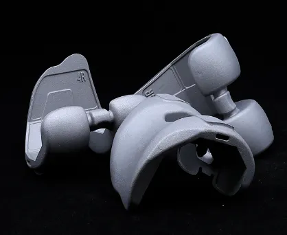
The femoral condyle is not only pivotal for joint mobility but also plays a significant role in maintaining knee stability. The shape and surface area of the condyles ensure proper distribution of forces across the knee, reducing excessive pressure on any single point. This distribution is vital in preventing premature wear of cartilage and the development of osteoarthritis.
From a biomechanical perspective, the two condyles have distinct roles. The medial condyle is generally larger and more weight-bearing, while the lateral condyle is smaller but crucial for facilitating knee rotation and lateral stability. Injuries or degeneration of either condyle can result in pain, joint instability, and impaired function, underscoring the importance of preserving the integrity of these structures.
Advanced imaging modalities such as MRI and CT scans allow clinicians to visualize the femoral condyle in great detail, facilitating early diagnosis of pathologies like cartilage defects, bone bruises, or fractures. Understanding the condylar anatomy also guides surgeons in planning procedures such as arthroscopy, osteotomy, or total knee arthroplasty, ensuring that treatments restore both the anatomical shape and biomechanical function of the knee.
The Role of the Lateral Epicondyle of Femur in Knee Mechanics
Situated just proximal and lateral to the lateral femoral condyle lies the lateral epicondyle of femur, a bony prominence that serves as a crucial attachment site for several soft tissue structures. Notably, the lateral collateral ligament (LCL) and the iliotibial (IT) band attach here, both of which contribute significantly to the lateral stability of the knee joint.
The lateral epicondyle of femur acts as a lever arm, facilitating the proper tensioning and mechanical advantage of these stabilizing structures. This function is especially critical during activities that involve side-to-side movements or sudden directional changes, such as sports or physically demanding work.
Injuries involving the lateral epicondyle of femur can vary from ligament sprains to avulsion fractures where the ligament or tendon pulls off a fragment of bone. Such injuries may lead to lateral knee pain, swelling, and instability, which can compromise mobility and quality of life if left untreated.
Chronic overuse or repetitive strain may also cause conditions such as lateral epicondylitis, although this term is more commonly associated with the elbow. Nonetheless, inflammation around the lateral epicondyle of femur can cause similar symptoms of pain and tenderness, requiring targeted rehabilitation.
Proper clinical evaluation, including physical examination and imaging, helps identify the extent of injury to the lateral epicondyle of femur. Treatment ranges from conservative management with physiotherapy and bracing to surgical repair in severe cases. Restoring the normal biomechanics of the lateral knee compartment is essential for preventing recurrent injuries and maintaining long-term joint health.
Weight Bearing Medial Femoral Condyle: Key to Load Distribution and Joint Longevity
Among the femoral condyles, the weight bearing medial femoral condyle holds a special significance due to its dominant role in supporting the body's weight during both static and dynamic activities. The anatomical axis of the lower limb passes slightly medial to the center of the knee joint, resulting in increased loading on the medial compartment.
This increased stress makes the weight bearing medial femoral condyle more susceptible to degenerative changes, particularly osteoarthritis. As cartilage thins and deteriorates under chronic load, patients often experience medial knee pain, stiffness, and reduced range of motion.
Subchondral bone beneath the cartilage of the weight bearing medial femoral condyle may also undergo cystic changes, sclerosis, or microfractures as a result of the altered biomechanics and inflammation. These pathological changes can exacerbate symptoms and accelerate joint degeneration.
Clinicians focus extensively on the medial femoral condyle when assessing patients with knee pain. Early detection of degeneration allows for interventions such as weight management, physical therapy to strengthen surrounding musculature, and joint unloading through orthotics or braces.
In advanced cases, surgical options like unicompartmental knee replacement target the weight bearing medial femoral condyle, replacing only the damaged portion while preserving healthy joint tissue. This approach can offer faster recovery and more natural joint kinematics compared to total knee replacement.
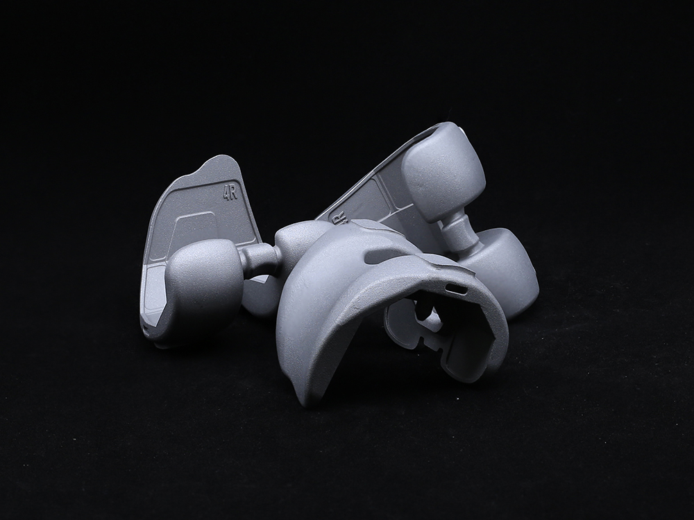
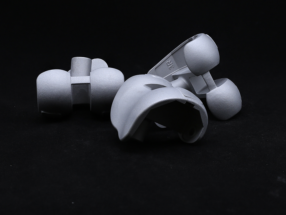
Recent advances in implant technology have led to the development of prostheses that closely replicate the shape and load-bearing characteristics of the weight bearing medial femoral condyle, optimizing outcomes and implant longevity.
Innovations in Treating Femoral Condyle Injuries and Degeneration
The evolving understanding of femoral condyle anatomy and biomechanics has fueled significant advancements in treatment options. For cartilage injuries involving the femoral condyle, procedures such as microfracture surgery, autologous chondrocyte implantation, and osteochondral grafting aim to regenerate cartilage and restore joint surface integrity.
When degenerative changes affect the weight bearing medial femoral condyle, orthopedic surgeons often consider joint-preserving surgeries such as high tibial osteotomy to realign the mechanical axis and offload the medial compartment. This can delay or prevent the need for knee replacement.
Total and partial knee arthroplasty procedures have seen improvements through patient-specific instrumentation and computer-assisted surgical planning. These technologies enhance the accuracy of implant positioning relative to the femoral condyle and lateral epicondyle of femur, improving knee mechanics post-surgery.
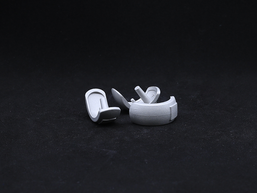
Moreover, cutting-edge materials like highly cross-linked polyethylene and cobalt-chromium alloys, as well as novel surface coatings, contribute to the durability and biocompatibility of knee implants. Such innovations are critical to replicating the natural function of the femoral condyle and maintaining joint health.
Rehabilitation techniques also emphasize restoring muscular balance around the knee, protecting the repaired or replaced femoral condyle, and retraining neuromuscular control to prevent future injuries.
femoral condyle FAQs
What is the role of the femoral condyle in knee joint function?
The femoral condyle forms the rounded ends of the femur that articulate with the tibia, allowing the knee to flex and extend smoothly. It supports weight and helps distribute forces evenly across the joint, reducing wear on cartilage.
Why is the lateral epicondyle of femur important in knee stability?
The lateral epicondyle of femur serves as an attachment for ligaments and tendons that stabilize the outer side of the knee, preventing excessive sideways movement and protecting the joint during dynamic activities.
What makes the weight bearing medial femoral condyle prone to osteoarthritis?
Because the weight bearing medial femoral condyle bears most of the body's load during activities like walking, it experiences higher stress. Over time, this can cause cartilage breakdown and degenerative changes leading to osteoarthritis.
How are injuries to the femoral condyle typically treated?
Treatment depends on injury severity and ranges from conservative care such as rest and physiotherapy to surgical interventions like cartilage repair or knee replacement to restore joint function.
Can knee implants effectively replicate the femoral condyle anatomy?
Yes, modern knee implants are designed to closely mimic the shape and biomechanics of the femoral condyle, ensuring natural movement and better distribution of forces post-surgery.
Get a Custom Solution!
Contact Us To Provide You With More Professional Services

