Understanding the Shallow Hip Socket
A shallow hip socket, medically termed acetabular dysplasia, is a condition where the hip socket or acetabulum does not fully cover the femoral head, leading to joint instability. This anatomical deficiency can be congenital or acquired and is a significant risk factor for early hip osteoarthritis and joint pain.
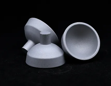
In a healthy hip, the acetabulum forms a deep, cup-shaped cavity that snugly encases the femoral head, facilitating smooth and stable ball-and-socket articulation. When this socket is shallow, the reduced coverage means that the femoral head has less support, making it more prone to subluxation (partial dislocation) or full dislocation during movement or weight-bearing activities.
Patients with a shallow hip socket often report symptoms such as groin pain, a feeling of hip “giving way,” stiffness, and difficulty performing activities that require hip stability like walking on uneven ground or climbing stairs. Left untreated, the abnormal mechanics increase stress on the labrum (the fibrocartilaginous rim) and cartilage, accelerating wear and degeneration.
Early diagnosis is vital. Physical examination combined with imaging techniques like X-rays, MRI, or CT scans allows physicians to assess the depth of the acetabulum and any associated damage. Advanced measurements such as the lateral center-edge angle help quantify socket coverage.
Treatment approaches depend on severity. Mild cases may benefit from physical therapy focusing on strengthening periarticular muscles to compensate for instability. In contrast, moderate to severe dysplasia often necessitates surgical intervention. Procedures like periacetabular osteotomy reshape and reorient the acetabulum to improve femoral head coverage, restoring joint stability and delaying osteoarthritis progression.
The advent of hip preservation surgery represents a major advancement for younger patients with shallow hip socket conditions, offering joint-saving options that can defer or even avoid total hip replacement.
Comprehensive Insights into Hip Socket Anatomy: Structure and Function
The hip socket anatomy is fundamental to the unique stability and mobility of the hip joint. The acetabulum, formed by the fusion of three pelvic bones—the ilium, ischium, and pubis—creates a deep, hemispherical cavity that articulates with the spherical femoral head.
Several key features define the hip socket anatomy:
The acetabular rim is reinforced by the labrum, a fibrocartilaginous ring that deepens the socket, improves joint congruity, and helps maintain the suction seal essential for joint stability.
The acetabular cartilage lining the socket is thickest in areas that bear the most weight, providing shock absorption and smooth movement.
The socket’s orientation in three planes (inclination, anteversion, and depth) governs how forces are transmitted through the joint during movement.
Disruption in any aspect of this anatomy—such as a shallow hip socket, labral tears, or cartilage degeneration—can compromise joint function and lead to pain and early arthritis.
Understanding the detailed hip socket anatomy enables orthopedic surgeons to tailor treatments. For example, in hip replacement surgery, replicating the natural socket shape and orientation is critical for implant longevity and restoring normal biomechanics.
Furthermore, the dynamic interplay between bone structure, cartilage, ligaments, and muscles around the hip socket anatomy forms the basis for comprehensive rehabilitation strategies aimed at preserving or restoring hip function.
Acetabular Cup Anteversion: Why Precise Implant Positioning Matters in Hip Replacement
In the realm of total hip arthroplasty (THA), the concept of acetabular cup anteversion is a cornerstone for ensuring the success and longevity of the prosthetic joint. Anteversion refers to the forward rotation or tilt of the acetabular component relative to the pelvis.
Correct acetabular cup anteversion allows the artificial femoral head to move smoothly within the socket, preventing impingement between components and reducing the risk of dislocation, which is one of the most common complications post-THA.
Surgeons aim for an anteversion angle typically between 15° and 25°, though this range may be adjusted based on patient-specific anatomy and activity levels. Too little anteversion increases posterior dislocation risk, whereas excessive anteversion predisposes to anterior dislocation and abnormal wear patterns.
Achieving optimal acetabular cup anteversion involves meticulous surgical planning supported by imaging studies like CT scans. Intraoperative navigation tools and robotic-assisted surgery have revolutionized accuracy in implant placement, helping surgeons maintain the ideal anteversion angle.
Beyond stability, proper anteversion influences the functional range of motion, helping patients return to daily activities and sports with confidence. It also minimizes asymmetric loading that can accelerate polyethylene wear or cause osteolysis around the implant.
Selecting the Ideal Acetabular Cup Size: Balancing Fit, Stability, and Longevity
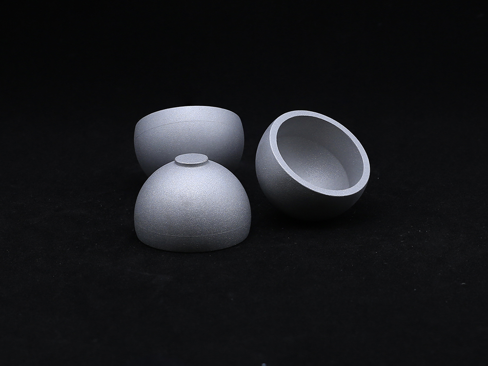
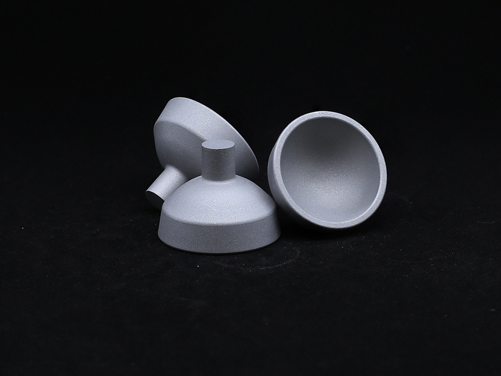
Choosing the correct acetabular cup size is as vital as positioning. The size must correspond closely to the patient’s acetabulum to ensure a secure press-fit and maximize bone-implant contact for biological fixation.
An undersized cup can lead to micromotion, early loosening, and instability, while an oversized cup risks fracturing the acetabular bone or causing impingement with surrounding tissues.
Modern implants offer a broad spectrum of sizes, often with modular liners to fine-tune the fit and optimize biomechanics. Preoperative templating using digital imaging allows surgeons to estimate the best acetabular cup size tailored to individual pelvic anatomy.
Bone quality is another consideration when selecting cup size. In patients with osteoporosis or acetabular defects, surgeons may augment fixation with screws or bone grafts to compensate for reduced bone stock.
The interplay between acetabular cup size and anteversion is critical to achieving stable, functional outcomes. Meticulous attention to these parameters reduces revision rates and improves patient satisfaction over the lifespan of the prosthesis.
Acetabular cup FAQs
What symptoms indicate a shallow hip socket?
Symptoms include groin or hip pain, a sensation of instability or “giving way,” stiffness, difficulty with weight-bearing activities, and sometimes audible clicking or catching during hip movement.
How is hip socket anatomy assessed clinically?
Assessment involves physical examination and imaging such as X-rays to measure socket depth and coverage, MRI to evaluate cartilage and labral integrity, and CT scans for detailed bone morphology.
Why is acetabular cup anteversion crucial in hip replacement?
Proper anteversion prevents prosthetic dislocation and impingement, ensuring smooth hip motion and implant durability. Incorrect anteversion is a leading cause of post-surgical complications.
Can acetabular cup size be adjusted during surgery?
Yes, surgeons often trial multiple sizes intraoperatively to achieve the best fit, sometimes using modular components to optimize stability and range of motion.
Is surgery the only option for treating a shallow hip socket?
No, mild cases may respond to conservative management like physical therapy and activity modification, but surgical correction is often needed to restore joint stability and prevent arthritis progression.
Get a Custom Solution!
Contact Us To Provide You With More Professional Services

